
Our History of Innovation
Pioneering Work
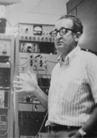
16 years before the founding of MBF Bioscience, the pioneering neuroscience researchers Dr. Edmund Glaser, MBF co-founder, and his colleague, Dr. Hendrik Van Der Loos create the first ever computerized microscope to help understand the brain.
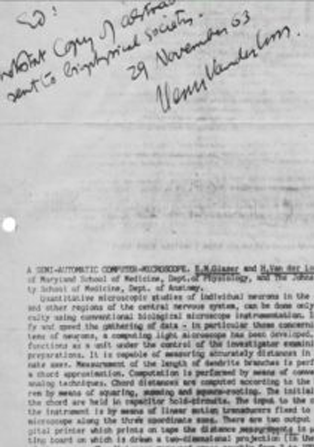
The origin of Digital Neuron Tracing and Reconstruction
The paper authored by Dr. Edmund Glaser and Dr. Hendrik Van Der Loos appears in IEEE Transactions on Biomedical Engineering titled “A semi quantitative computer-microscope for the analysis of neuronal morphometry.” This paper presciently describes the importance of quantitative neuron reconstruction and is the seminal work that eventually led to the Neurolucida system.
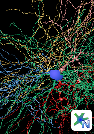
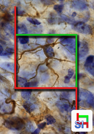
Neurolucida
Neurolucida, the culmination of over 25 years of pioneering experience in neuroimaging by the father and son team of Dr. Edmund Glaser and Jack Glaser, is invented. This system remains the gold standard for neuron tracing. It is used in over a thousand labs and is the most cited neuron tracing program in the world with over 6,000 citations. It has been continuously developed for over 30 years. Neurolucida, is the most widely used Neuron Tracing and Brain Mapping system in research laboratories throughout the world.
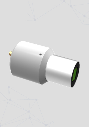
Lucivid: Minature Microscopy Display Technology
Lucivid, an innovative microdisplay used by researchers to trace neurons, perform stereology, and stimulate retinal cells, is introduced. For neuron tracing and stereology, the Lucivid was used instead of a camera to gather data while looking directly though the microscope oculars at your specimen. It superimposes what is displayed on your computer monitor into the microscope's field-of-view so you can trace neurons and mark cells for quantification.
Stereo Investigator
Stereo Investigator, the leading software and system for unbiased stereology, is invented. It is still in continuous development evolving to meet the most exacting quantification needs of scientists. Stereo Investigator is now the world’s most cited stereology system with over 8,000 citations.
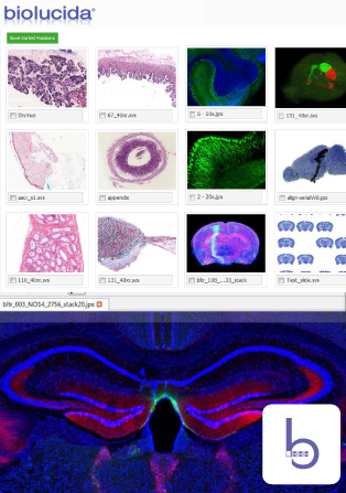
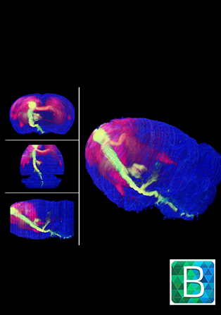
Biolucida - Big Image Data Sharing and Collaboration
With two different specialized editions for Medical Education and Research, Biolucida allows scientists to easily view data sets that have been too voluminous and complex for traditional systems in the past to be viewed with ease. More than 100,000 medical students have learned pathology and histopathology with Biolucida. One resource, the American Association of Anatomists' Virtual Microscopy Database, holds over 3,300 different slides from 15 different institutions are available for free for professors teaching histology and histopathology.
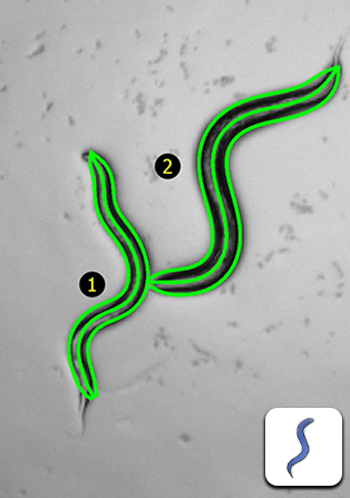
WormLab - C. elegans Behavioral Analysis Made Easy
WormLab, software and hardware for imaging, tracking, and analyzing C. elegans and other worms, is invented. C. elegans is used in multiple research fields such as neurodegeneration, genetics, aging, development, and even toxicology.

Vidrio Technologies
Vidro Technologies, now a part of MBF Bioscience family, was incubated at the Janelia Research Campus to provide imaging solutions for neuroscience. It went on to launch ScanImage, vDAQ and the RMR.
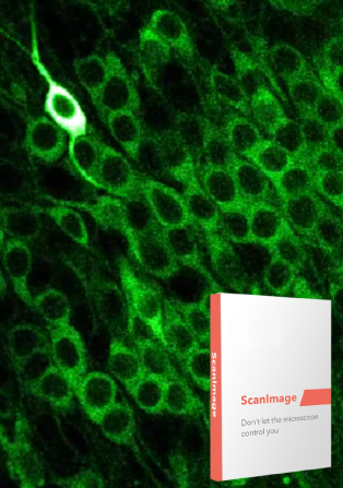
ScanImage - Software for Controlling Multiphoton Microscopes
Don’t let the microscope control you! Originally developed in neuroscience labs by Drs. Karel Svoboda and Vijay Iyer in 2003. Then in 2014, Vidrio Technologies, now a part of MBF Bioscience, extended the capabilities of this popular lab developed software package and released a commercial version. ScanImage is the most widely used software for controlling multi-photon and laser scanning microscopes.
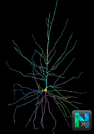
Neurolucida 360 - Automatic 3D Neuron Reconstruction
Neurolucida 360 is the premier tool for 3D automatic neuron reconstruction used by scientists and researchers; allowing them complete their research as quickly as the speed of neuroscience. This software allows you to automatically reconstruct neurons of all sizes. Used in laboratories worldwide. Neurolucida 360 is the leading software for automatic neuron tracing.
BrainMaker - Reconstructing the Brain
BrainMaker, the fast and smart software that automatically creates 3D reconstructions of the brain from serial sections of whole slide images. Even if you mounted a section upside down, no worries, BrainMaker can fix that in the alignment process. This software can assist you with cell mapping, cytoarchitectonics and other areas that require the characterization of neuronal circuitry to create a comprehensive anatomical reference.
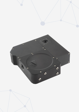
Rapid Multi Region Scanner (RMR)
The RMR Scanner combines the flexibility of galvo mirrors with the speed of resonant scanning for 2 photon imaging. Its novel design combines two galvo and one resonant mirror into one compact device. The scanner is fully compatible with ScanImage's®powerful scanning modes. The flexible mounting options allow installation on a Thorlabs BScope, Sutter MOM and DIY microscopes.
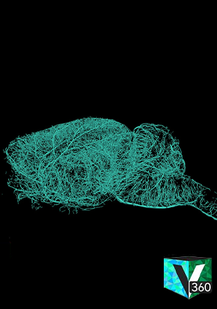
Vesselucida 360 - Automatic Reconstruction of Blood Vessels and Microvasculature
Vesselucida 360 provides groundbreaking software for researchers who are studying neuroscience, cancer, diabetes, strokes, and other conditions that affect microvasculature. Quickly segment and fully reconstruct vessels and microvasculature in 3D to obtain reliable data about both vessels and microvessels.
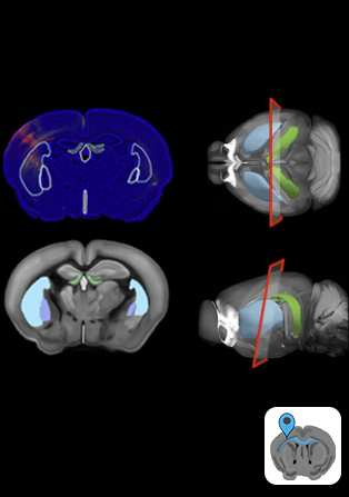
NeuroInfo – Atlas-based, Brain-Wide Screenings for Cells and Biomarkers
Perform brain-wide analyses of cells and biochemical markers with NeuroInfo. Sophisticated detections using advanced, deep learning methods. You no longer have to manually click cells or hand draw your anatomic boundaries because NeuroInfo will automatically categorize them into neuroanatomical regions.
Stereo Investigator - Cleared Tissue Edition: A New Gold Standard in Unbiased Stereology
This software edition is specially engineered to allow researchers to use the world’s most cited unbiased stereology software to count cells and analyze fibers in intact, cleared tissue specimens imaged with light sheet or confocal microscopes. It even works with multi-terabyte 3D images.
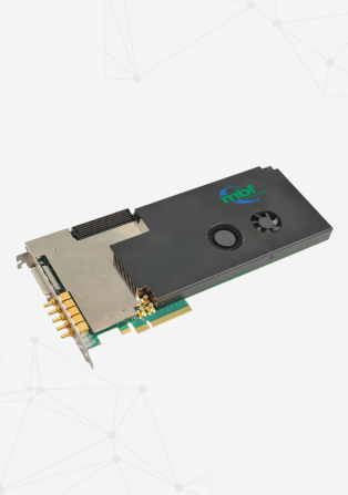
vDAQ - Data Acquisition Board for Multiphoton and Laser Scanning Microscope Control
Tightly integrated with ScanImage, vDAQ controls galvos, resonant scanners, pockels cells, piezo objective positioners, shutters and much more. It greatly simplifies the wiring complexity of microscopes by eliminating the need for additional 3rd party DAQ hardware.
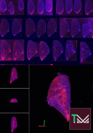
TissueMaker - Create 3D Reconstructions of an Entire 3D Organ
Turn a series of serial sections imaged with whole slide scanners into a 3D reconstruction of any organ.
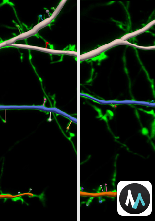
MicroDynamix - Elucidating and Analyzing Dendritic Spine Changes
MicroDynamix expands upon Neurolucida 360's dendritic spine detection and reconstruction by providing new functionality for dynamic in vivo analyses in time series from repeated imaging experiments. You can automatically align the images in 3D, reconstruct dendritic branches, detect dendritic spines, and identify important metrics like length, luminance, classification, and more for accurate comparison between dendritic spines.
MicroFile+ Helps Standardize and Work with Large Microscope Image Files
To solve a problem faced across laboratories and imaging facilities, MicroFile+ was introduced to efficiently convert 2D and 3D images from almost any source and format into manageable, standardized experimental results. With this, you can organize, store, and analyze big data by converting the data into compact, easy to manage files that can be instantly loaded by MBF Bioscience technologies for analysis. Nearly any type of image, along with its metadata, can be converted to the jpeg20000 or OME Tiff standards.
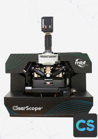
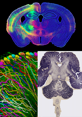
ClearScope – Revolutionary Light Sheet Microscope Even for Very Large Specimens
The revolutionary light-sheet theta microscope, ClearScope, was created in collaboration between Columbia University and MBF Bioscience. With the ability to image large specimens, even tissue slabs from human brains, and small specimens, such as whole mouse and rat brains, ClearScope uniquely meets any challenge researchers may face when conducting light-sheet microscopy research.

Vidrio joins the MBF family
Recognizing the emergence and importance of multi-photon imaging and advanced solutions, MBF adds Vidrio Technologies of Virginia to its growing family of bioscience products. The powerful combination of these two companies sets MBF on an accelerated path towards new and exciting products for the future, and adds intellectual property and valuable employees to MBF’s overall offering.
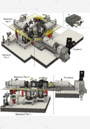
SLAP2 Kit - Unparalleled Optical Imaging of Neuronal Activity
A revolutionary new microscope kit based on the technological breakthrough called Scanned Line Angular Projection (SLAP) two-photon laser scanning microscopy. This microscope was invented by Dr. Kaspar Podgorski. The game-changing innovation in the SLAP2 is the ability to perform unparalleled optical imaging of neuronal activity in populations of neurons at subcellular spatial resolution and temporal resolution in the millisecond range both in vivo and in vitro using a variety of fluorescent indicators.
Neurophotometrics joins MBF family
MBF Bioscience has added Neurophotometrics, the leading fiber photometry company, to our cutting-edge bioscience lineup. Both brands share a strong commitment to helping scientists bring new insights to the future of bioscience. Our teams are excited to combine our efforts together to develop new products and bridge technologies, propelling the advancement of bioscience.