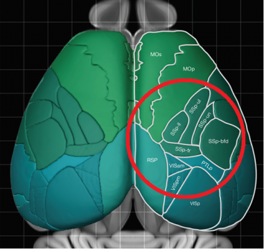
2-photon microscopy has been established as a standard tool for recording neuronal activity in live, awake animals. Until recently, there has been a tradeoff: image a small set of neurons with cellular resolution, or image larger brain regions while sacrificing resolution. In 2016, a 2-Photon Random Access Microscope (2PRAM) was developed in the Svoboda lab at HHMI’s Janelia Research Campus [1]. This microscope combines for the first time, a large field of view (∅ 5 mm x 1 mm cylinder) with sub-cellular resolution.
ScanImage was developed in close collaboration with the Svoboda lab to enable mesoscale imaging, and it is the only software that can operate the mesoscope. ScanImage’s flexible multi-region of interest (mROI) feature allows either imaging the entire 5mm field of view, or select multiple smaller regions. By using the mesocope’s remote focusing system, the ROIs can be extended into 3D volumes. To compensate for the objective’s inherent field curvature, ScanImage adjusts the focus via the remote focusing system depending on the location of the scanned region.
More information about the mesoscope design can be found on the Janelia website and the Thorlabs product page.
[1] Sofroniew, N. J., Flickinger, D., King, J., & Svoboda, K. (2016). A large field of view two-photon mesoscope with subcellular resolution for in vivo imaging. Elife, 5, e14472.
